INTRODUCTION
In 1969, Branemark et al1 published landmark research documenting the successful osseointegration of endosseous titanium implants. Since then, these methods for surgical placement of dental implants had a profound influence on
the practice of dentistry. For the success and longevity of dental implant osseointegration is very important. To prevent the failure in implants stress should be evaluated at the implant bone interface. Most common complication in an implant reconstruction is related to occlusal overload and stress related factors. Excess stresses to an implant interface may cause early to late implant failures, crestal bone loss, porcelain fractures, unretained restorations, implant component failure and screw loosening.
CAUSES OF OVERLOADING
a. Overloading Factors related to Patient
b. Overloading Factors related to the Implants An implant team should evaluate more than 60 force factors before developing a treatment plan. Some force factors are more important than others. So these forces should be evaluated in the
(1) Magnitude, (2) Duration (3) Type (4) Direction (5) Multiplication Factors.2
Several factors may multiply or increase the effect of the other conditions.
Dental conditions primarily include the following:
1) Parafunction
i) Bruxism
ii) Clenching
iii) Tongue Thrust
2. Crown Height
3. Masticatory Dynamics
4. Position of the Abutment in the Arch
5. Direction of Load
6. Nature of the Opposing Arch
1) Parafunctional Forces Parafunctional forces on teeth or implants are characterized by repeated or sustained occlusion
and have long been recognized as harmful to the stomatognathic system. The most common cause of implant failure after successful surgical fixation or early loss of rigid fixation during the first year of implant loading is the result of parafunction. Such complications occur with greater frequency in the maxilla, because of a decrease in bone
density and an increase in the moment of force. 3 The parafunctional groups presented in this chapter are divided into bruxism, clenching, and tongue thrust or size of the tongue.
A) Bruxism - The forces involved are in significant excess of norma1 physiologic masticatory loads. Bruxism may generate several hours per day of increased force on the teeth. A 37-year-old patient with a long history of bruxism recorded a maximum bite force more than 990 psi (4 to 7 times normal).
4B) Tongue Thrust- is the unnatural force of the tongue against the teeth during swallowing.5A force of approximately 41 to 709 g/cm2 on the anterior and lateral areas of the palate has been recorded during swallowing. A tongue thrust habit may lead to tooth movement or mobility, which is especially of consequence when implants are present in the same
quadrant.
1) Crown Height
The crown height is the vertical cantilever or the lever. The greater the crown height, greater the movement of force under lateral loads. Because the stresses are concentrated at the crest of rigidly fixated implant, the crown height multiplier increases stress rapidly. For every 1mm crown height increase, force increase may be 20%. Therefore the crown
height increased from 10-20% may increase the stress by 200%. As the bone resorbs, the crown height become larger, but the available bone height decreases. An indirect relationship is found between the crown and implant height which magnifies the stresses.
 | Fig. 1) Bone resortion occurs at the crest, increases the crown height results in more stresses.
 |
These are responsible for the amount of force exerted on implant system. The dentist evaluates the several conditions under this heading: patient size, sex, age and skeletal position. 6 The size of the patient can influence the amount of bite force. The larger athlete men generate greater forces; than patient of weak physical condition.
2) Position within the Arch
Maximum bite forces in the anterior incisor region range from 35 to 50 psi; those in the canine region range from 47 to 100 psi and those in the molar area range from 127 to 250 psi, Mansour et al 7 in addition, the force at the second molar was 10% higher than at the first molar, indicative range from 140 to 275 psi.
3) Direction of Load
The direction of the occlusal load results in significant difference in the amount of force exerted on an implant. Forces are tensile, compressive, or shear to the implants. Bone is strongest to compressive forces, 30% weaker to tensile loads, and 65% weaker to shear loads; three dimensional stress analyses has shown that almost all the stresses occur in
the coronal half of the implant bone interface. Much less stress occurs with vertical loads compared with the angled load on implant. Lateral forces represent the 50 -200% increase in stress compression as compared with the vertical loading.
4) Opposing Arch
Natural teeth transmit greater impact forces through occlusal contacts than do soft tissueborne complete dentures. In addition maximum occlusal force with complete dentures is reduced and may range from 5 to 26 psi. Muscle atrophy, thinning of the oral tissues with age or disease, and bone atrophy often occurs in the edentulous patient as a function of time. Complete implant fixed prosthesis does not benefit from proprioception as do natural teeth. And patients bite with a force four times greater than with natural teeth.
STRESS FACTORS RELATED TO IMPLANTS
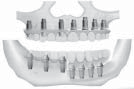
Available bone describes the amount of bone in the edentulous area considered for implantation. The amount of bone is measured in width, height, length, angulations, and crown height, implant body ratio for proper size of the Implant. Improper selection of implants in relation to available bone increases the stresses at the implant bone interface.
1) Size of the Implant
A) Implant Length-The Implant length
corresponds to the height of available bone. Increased implant length is usually not significant at the crestal bone interface, but is beneficial for initial stability and overall amount of bone implant interface. Increased length also provides resistance to torque or shear forces. An Implant 3 mm longer provides more than 10 % increase in surface area. This
increased length does little to decrease the stress at Transosteal region around implant at crest of the ridge.8
B) Implant Width-The surface area of each implant is directly related to the width of the implant. Each 0.25 mm of implant diameter, the surface area increases by the 5% -8%. Past theories suggested that the implant height is more important than the width, but it is not true as occlusal load concentrates most at the crest of the bone, so implant width is more
important.9
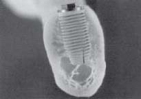
2) Implant Design
Threads are designed to maximize the initial contact, enhance surface area and facilitate the dissipation of stresses at the bone implant interface. Functional surface area per unit length of the implant may be modified by varying three thread geometry parameters: Thread Pitch, Thread Shape and Thread Depth.
A) Thread Number- A threaded implant with 10 threads for 10mm has more surface area than one with 5 threads.
B) Thread Depth- A thread depth of 0.2 mm has less surface area than-an implant with 0.4 mm.
C) Thread Shape- The thread shape is an important characteristic of overall thread geometry it include square, v shaped, buttress geometry. The shear force on v thread face is about 10 times greater than the shear force on square thread.
 | Fig 3) A) Two number abutments results in more forces. B) Three abutment reduces the stress transmission to bone.
|
3) Abutment Number
The overall stress on the implant system may be reduced by increasing the number of abutment over which the force is applied. The most effective method to increase the surface area of implant support is by increasing the number of implants used to support prosthesis. The force distributed over three abutments resulted in less stress to crestal bone than two abutments.
 | Fig 2) Increased number of Threads provides more surface area for less dense bone
|
4) Abutment Position
Implant positioning also is related to implant number because more than two implants are needed to form a biomechanical tripod, that is, not a straight line. The suggestion is that multiple units be placed in a staggered buccal
Abutment offset (tripod effect). Cantilevers are force magnifier and represent a considerable risk factor. Therefore implant
number and position should aim at eliminating cantilevers ‘whenever possible, especially when other force factors are increased.
SUMMARY
CONCLUSION
Complications and loss of implants can be costly, both in terms of time and financial resources. Loss of integration can be troublesome, resulting in an edentulous space more difficult to restore than prior to implant placement. The ability
to reliably identify patients and conditions with greater potential for success would be valuable
REFERENCES
1. Melanie RW and Stanley GV, A review of selected dental literature on evidence based treatment planning for dental
implants: Report of the Committee on Research in Fixed Prosthodontics of the Academy of Fixed Prosthodontics, J
Prosthet Dent 2004; 92:447-62.
2. Bidez MW, Misch CE: Force transfer in implant dentistry basic concepts and principles, Oral Implantol 18:264-274,
1992.
3. Jaffin R, Berman C: The excessive loss of Branemark fixtures in type V bone: a 5 year analysis, J Periodontol62: 2-4, 1991
4. Gibbs CH, Mahan PE, Mauderli A, et al: Limits of human bite force, J Prosthet Dent 56:226-229, J986.
5. Kydd WL, Toda JM: Tongue pressures exerted on the hard palate during swallowing, Am Dent Assoc 65:319, 1962
6. Howell AH, Bruderold F: Vertical forces used during chewing of food, J Dent Res 29:133, 1950
7. Mansour RM, Reynik RJ, and Larson PC: Piezoelectric transducers for biting force measurements! PhD thesis. In
Abstracts of the twenty-seventh ACEMB, 1974.
8. Weinberg LA, Kruger B: An evaluation of torque on implant/ prosthesis with staggered buccal and lingual off- set, Int J
OralMaxillofacImplants16:253, 1996.
9. Sertgoz A, Guvener S: Finite element analysis of the effect of cantilever and implant length on stress distribution on implant supported prosthesis. J Prosthet Dent 165-169, 1996. |

